The Mueller Weiss syndrome is a rare cause of pain within the midfoot in adults that has been less commonly called Brailsford disease. Mueller Weiss Syndrome is a spontaneous onset osteonecrosis of the navicular within the foot. You will find there’s a much more well-known problem of the navicular in children referred to as Köhler disease, which is also an osteonecrosis in the tarsal navicular bone, but they are totally different disorders because of the character with the developing bone tissues in children. The disorder was first reported by Schmidt back in 1925. It had been W Muller who afterwards proposed how the underlying mechanism with the disorder has been resulting from an excessive compression force on the mid-foot region. About the same period, K Weiss, documented which the appearances on radiographs were similar to those seen in a disease called Kienbock disease, and this is an osteonecrosis. Those two accounts resulted in the most commonly used name for this problem, Mueller Weiss syndrome.
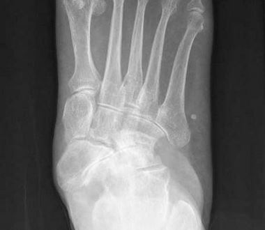
Mueller Weiss disease commonly affects adults between forty and sixty years of age (Köhler disease has a common starting point around five years of age). Mueller Weiss Syndrome is more common in females. It can affect just one foot, or it may affect both feet. The traditional symptoms include the progressive oncoming of discomfort in the midfoot and hindfoot which could often be localised to the most painful location being about the navicular bone. A flat foot is in addition more prevalent in those with this problem. The easiest way to diagnose Mueller Weiss disease is by using imaging. On x-ray there will seem like a collapse of portions of the navicular and a whiteness with comma-shaped deformity of the lateral aspect. A CT scan could also present similar irregularities and can be used to appraise the stage with the condition in additional detail. A magnetic resonance image is usually a lot more sensitive to help with the verification because it is able to find alterations in the bone marrow.
Mueller Weiss disorder is usually progressive and may produce significant pain and be fairly debilitating, so treatment needs to be started as early as possible in order to avoid the navicular bone from being impaired too much. Initial treatment methods are to reduce weight bearing, maybe some pain relief drugs and make use of supporting shoes or boots. Generally foot supports are widely-used to help further stabilise the bones and support the mid-foot (arch) of the feet. This will keep a lot of load off the navicular bone. If that’s not necessarily making a difference, then a more limitation in exercise levels is needed so there is a lesser amount of strain about the painful region. A moon boot or walking splint is usually the next phase to help protect and also immobilise the region if the symptoms are not improving. If all of these conservative techniques usually do not help, you can also find surgical alternatives that can help with the soreness however can often leave a little minor disability, which can be a lot better than the persistent pain of an active disease. The actual surgical treatment might be a decompression of the navicular bone with drilling. Another option in case there are areas of bone deterioration are a operative fusion of the important joints about the damaged bone..
Advertisement:

- Hardcover Book
- Wong-Chung, John (Author)

- Audible Audiobook
- Yella A. Deeken (Author) - Stephan Müller (Narrator)

- Amazon Kindle Edition
- Müller, Dr. Maria (Author)
I get commissions for purchases made through links on this website. As an Amazon Associate I earn from qualifying purchases.


1 comment
Nadal's Foot Injury from Tennis | Curation of Knowledge
Feb 20, 2022
[…] foot injury that Nadal has was a condition known as Müller-Weiss Syndrome or disease. It is a pretty uncommon spontaneous necrosis or loss of supply of blood for the navicular bone in […]